Wednesday, April 20, 2011

Elvis had a Pelvis with...


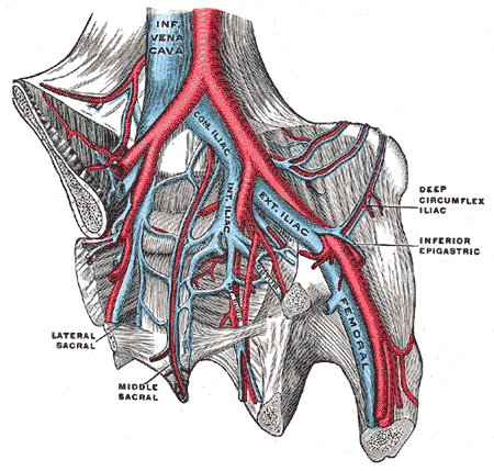
The pelvis is the region of the body intermediate to the abdominal cavity and the lower limbs, including the pelvic girdle.  The pelvic girdle surrounds the pelvis and is part of the appendicular skeleton.  The pelvic girdle is made up of the right coxal bone, left coxal bone, and the sacrum.  Also made up of three bones is the coxal bone, the most lateral aspects of the pelvic girdle, formed by the fusion of the ilium, ischium, and pubis.  Formed by the fusion of five sacral vertebrae is the sacrum, the most posterior portion of the pelvic girdle.  The most anterior aspect of the pelvic girdle is the pubic symphysis, joining the two coxal bones.


Although there are similarities among pelvic girdles, not all pelvic girdles are made equal.  The male and female pelvic girdles, as a particular focus, are different in many ways and these differences are typically used when identifying the sex of skeletal remains.  The general structure is thick and heavy in males, but thin and light in females.  Superior to the pelvic inlet is the greater pelvis which is deep in males, yet shallow in females.  Another difference is seen in the lesser pelvis, the region between the pelvic inlet and pelvic outlet, which is narrow and deep with a tapering shape in males and is wide and shallow with a cylindrical shape in females.  Also, the pelvic inlet is heart-shaped and narrow in males, leading to a relatively small pelvic outlet while the female pelvic inlet is oval-shaped and wide in females.

The differences between the male and female pelvic girdles are numerous due to the child-bearing function in females.  The pubic arch is the space between the junction of the inferior rami of the iscium and pubis on the left side and the same on the right side.  Expectedly so, the pubic arch of the male is narrower than that of the female as is the greater sciatic notch.  Also, the obturator foramen is of a round shape in males and oval in females.  Finally, in terms of the acetabulum, it is larger in males than in females.  Some of the differences between the pelvic girdles of males and females, as with the greater pelvis, can be attributed to the greater muscle mass of males in relation to females.
 

Within the lesser pelvis of both males and females is the pelvic diaphragm, separating the pelvic cavity from the perineum.  Forming the pelvic floor, the pelvic diaphragm consist of the coccygeus and levator ani muscles and their associated fascia.  Making up the larger portion of the pelvic floor is the levator ani, which is made up of three parts:  the puborectalis, the pubococcygeus, and the iliococcygeus.  The puborectalis forms a sling around the posterior aspect of the anorectal junction and aids in retaining fecal continence.  The levator ani function together to support the viscera with tonic contraction and to aid in forced expiration, among other activities, with active contraction.  Relaxation of the levator ani occurs for urination and defecation to occur.
The perineum, which includes the anus and external genitalia, and has additional muscles associated with the external genitalia.  Such muscles can be found within the pelvic cavity and include the ischiocavernosus and bulbospongiosus muscles.   The ischiocavernosus muscle acts to compress the corpus cavernosum.  The bulbospongiosus muscle has different actions in males and females.  In males, the bulbospongiosus muscle acts to compress the bulb of the penis and also compresses the spongey urethra.  In females, the bulbospongiosus muscle acts to compress the vestibular bulb and constrict the vaginal orifice.

There are several ligaments associated with the pelvis.  Females have a ligament called the suspensory ligament of the ovary that is a fold of peritoneum through which the ovarian vessels, lymphatics, and nerves pass to cross the pelvic brim.  To an extent, the suspensory ligament is continuous with the broad ligament.  The broad ligament serves to anchor the uterus in place, extending from the uterus to the pelvic floor and the walls of the pelvis.  Another ligament that serves to anchor the uterus is the cardinal ligament which maintains the anteverted position of the uterus.  During pregnancy, the round ligament maintains the anteverted uterine position rather than the cardinal ligament.

A. Robinson
Sources:
Moore, Dalley, Agur: Clinically Oriented Anatomy. 6th ed. Baltimore:  Lippincott Williams & Wilkins, 2010.

Tuesday, April 19, 2011

SVC & IVC

The superior vena cava (SVC) and the inferior vena cava (IVC), the largest veins in the human body, are part of the systemic circulation.  The SVC and IVC return deoxygenated blood from structures superior (except the lungs and heart) and inferior to the diaphragm, respectively, to the right atrium of the heart.  Formed by the junction of the left and right brachiocephalic veins, the SVC extends only a short distance to reach the right atrium.  The IVC is formed by the union of the left and right common iliac veins at the level of L5.  Extending from L5, the IVC passes through the vena caval foramen of the diaphragm to empty into the right atrium of the heart.  The IVC is greater in length than the SVC and has significantly more tributaries draining directly into it.

Due to the brachiocephalic veins being formed by the union of the internal jugular veins and the subclavian veins, the left and right brachiocephalic veins drain deoxygenated blood from the head, neck, and upper limbs into the SVC.  The right brachiocephalic vein descends inferiorly, but the left brachiocephalic vein passes at an angle anterior to the branches of the aortic arch.  The difference in paths of the left and right brachiocephalic veins is due to the position of the SVC being on the right side of the superior mediastinum just to the right of the trachea.  The left brachiocephalic vein is over twice as long as the right brachiocephalic vein due to its extended course.  The left brachiocephalic vein not only crosses to the right side of the superior mediastinum, but it must clear the branches of the aortic arch.  Formed by the position of the portion of the SVC within the middle mediastinum relative to the ascending aorta is the posterior boundary of the transverse pericardial sinus.

The azygos vein is unique in that it drains directly into the SVC whereas the left and right brachiocephalic veins join to form the SVC.  The azygos vein carries deoxygenated blood from the thoracic cage. 

The common iliac veins being formed by the union of the internal and external iliac veins.  The internal iliac vein receives deoxygenated blood from the gluteal vein, internal pudendal vein, obturator veins, lateral sacral veins, middle hemorrhoidal vein, vesical vein, uterine vein, and vaginal veins.  In contrast to the many tributaries of the internal iliac vein, the external iliac vein receives the deoxygenated blood from the femoral vein, deep circumflex iliac vein, and the inferior epigastric vein.  Keeping these contributing veins in mind, it is easier to understand that the common iliac veins are moving deoxygenated blood from the gluteal region, external genitalia, pelvic structures, and lower limbs into the IVC.  The IVC lies to the right of the abdominal aorta as it ascends retroperitoneally along the right side of the vertebral column.  Differences in drainage occur due to the IVC being located on the right side of the body.


Draining directly into the IVC are, from most inferior to most superior, the lumbar veins, ovarian/testicular vein, renal veins, suprarenal vein, hepatic veins, and the inferior phrenic vein.  One of the differences in drainage into the IVC occurs with the ovarian/testicular veins.  The ovarian/testicular vein on the right side drains directly into the IVC, but ovarian/testicular vein on the left side drains into the left renal vein.  Another difference in drainage into the IVC occurs with the suprarenal veins.  The right suprarenal vein drains directly into the IVC, but the left suprarenal vein drains into the left renal vein.  The deoxygenated blood is drained to the right atrium, where it enters through a semilunar valve which prevents backflow of blood.

There are lumbar veins on each side of the IVC.  The lumbar veins receive blood from the epigastric veins, vertebral plexuses, as well as other smaller tributaries.  The left lumbar veins are longer than the right because the IVC is on the right side of the vertebral column and because they pass behind the abdominal aorta.  The renal veins are large and pass in front of the renal arteries.  Passing in front of the abdominal aorta, the left renal vein is longer than the right renal vein and empties into the IVC just above the level of the right renal vein.  Due to the differences in drainage previously mentioned the left testicular/ovarian vein, the left inferior phrenic veins, and the left suprarenal vein.

The hepatic veins receive deoxygenated blood from the central veins of the liver.  There are two groups that make up the hepatic veins:  the upper group and the lower group.  The veins of the lower group tend to be smaller than those of the upper group.  Lastly, are the inferior phrenic veins which receive deoxygenated blood from the diaphragm.  The right inferior phrenic vein empties into the IVC, but the left inferior phrenic vein is sometimes seen with two branches.  One of the branches passes through the diaphragm and then empties into the IVC and the other branch empties into either the left renal or the left suprarenal vein.

A. Robinson
Sources:

Monday, April 18, 2011

Aortic

The aorta carries oxygenated blood to the body and originates at the aortic orifice from the left ventricle of the heart.  From the aorta's point of origin to the arch of the aorta is referred to as the ascending aorta.  The aortic arch begins at the level of the sternal angle and rises anterior to the right pulmonary artery, reaching its highest point to the left of the trachea, and descending posterior to the left root of the lung.  Posterior to the second left sternocostal joint, the aortic arch continues downward as the descending aorta, also called the thoracic aorta.  Once the thoracic aorta passes through the aortic hiatus of the diaphragm on its downward journey, it becomes the abdominal aorta.  The abdominal aorta reaches a termination point just inferior to the umbilicus and slightly to the left of the midsagittal plane as it bifurcates into the left and right common iliac arteries.

The only branches of the ascending aorta are the left and right coronary arteries.  As the aorta continues, there are three branches from the aortic arch.  In order from the most proximal in relation to the ascending aorta, the branches of the aortic arch are the brachiocephalic trunk, the left common carotid, and the left subclavian arteries.  Continuing inferiorly, the thoracic aorta gives rise to four pairs of branches and three unpaired branches.  As the thoracic aorta descends, there are the paired bronchial, posterior intercostal, subcostal, and superior phrenic arteries.  Also found as the thoracic aorta descends are the unpaired mediastinal arteries, esophageal arteries, and pericardial arteries.  The unpaired arteries of the thoracic aorta are not paired with their partner branches, but there are more than one of each of these arteries coming off of the thoracic aorta.  After passing through the aortic hiatus, there are four unpaired branches and five paired branches from the abdominal aorta.  The unpaired branches from most superior to most inferior are the celiac trunk, superior mesenteric artery, inferior mesenteric artery, and the median sacral artery.  Also found along the abdominal aorta's descent are the paired branches of the inferior phrenic, middle suprarenal, renal, ovarian/testicular, and lumbar arteries. 

Aorta - Anatomy : Ascending aorta, Aortic bulb, Aortic arch, Aortic isthmus


The branches from the ascending aorta have several significant branches of their own.  From its point of origin, the left coronary artery (LCA) courses between the left auricle and the left side of the pulmonary trunk and through the coronary sulcus where at the superior margin of the anterior IV groove it divides into the anterior IV branch and the circumflex branch.  In some people the anterior IV branch gives off a lateral branch which courses over the anterior surface of the heart.  The branches of the LCA also include a left marginal artery and sometimes the nodal branch to the SA node.  On the other hand, the RCA runs to the right side of the pulmonary trunk through the coronary sulcus and typically gives off a branch to the SA node, the SA nodal branch.  The RCA continues through the coronary sulcus and gives off a second branch, the right marginal branch; a third branch, the atrioventricular nodal branch; and usually gives off the posterior IV branch as well.

The branches of the aortic arch also have their own significant branches.  The brachiocephalic trunk bifurcates into the right common carotid artery and the right subclavian artery.  Dividing in the cervical region at the fourth cervical vertebrae, the left common carotid artery forms the external and internal carotid arteries of the left side.  While not having branches in the mediastinal region, the left subclavian artery does branch outside of this area to give rise to the vertebral artery, internal thoracic artery, thyrocervical trunk, costocervical trunk, and the dorsal scapular artery.  Once the left subclavian artery reaches the lateral border of the first rib it is referred to as the axillary artery.  It is important to note that variation in regards to the circulatory system in some form is far more common than one might expect and should always be considered.  The aortic arch branching pattern of the brachiocephalic trunk, left common carotid, and the left subclavian is referred to as the "Type A" branching pattern.

In regards to the branches of the thoracic aorta, the bronchial arteries generally run along, and branch with, the bronchi to supply the lungs with oxygenated blood.  The subcostal arteries have a posterior branch that is significant, but otherwise the subcostal arteries, posterior intercostal, and superior phrenic arteries give rise to small vessels that are unnamed and serve to distribute successively smaller vessels to supply oxygenated blood to tissue.  The subcostal arteries supply oxygenated blood primarily to the upper, posterior abdominal wall while the posterior intercostal artery branches supply the thoracic wall.  The vessels of the superior phrenic arteries supply the posterior region of the superior surface of the diaphragm.  The mediastinal arteries are once again small vessels that distribute oxygenated blood to tissue, in this case mostly the lymph glands and tissue of the posterior mediastinum.  The esophageal and pericardial arteries supply oxygenated blood through successively smaller vessels to the esophagus and pericardium, respectively.

Several of the branches of the abdominal have their own significant branches.  The celiac trunk has three main branches which are the left gastric artery, the common hepatic artery, and the splenic artery.  The superior mesenteric artery also has several main branches which are the inferior pancreatoduodenal artery, middle colic artery, right colic artery, intestinal arteries, and the ileocolic artery.  Somewhat similar to the superior mesenteric artery in its subsequent branches is the inferior mesenteric artery.  These subsequent branches are the left colic artery, sigmoid branches, and the superior rectal artery.  Of a somewhat lesser divisive status are the inferior phrenic arteries which both divide into a medial and lateral branch and give rise to the superior suprarenal branches.  The renal arteries serve their purpose of supplying the kidneys with oxygenated blood without giving rise to branches for other purposes.  Serving the purpose of sending successively smaller branches to supply oxygenated blood to certain areas without any major branches are the median sacral artery, the middle suprarenal arteries, and the lumbar arteries.

A. Robinson
Sources:
Moore, Dalley, Agur: Clinically Oriented Anatomy. 6th ed. Baltimore:  Lippincott Williams & Wilkins, 2010.
http://www.clevelandclinic.org/heartcenter/pub/guide/heartworks/bloodvesselpics.htm
http://www.imaios.com/en/e-Anatomy/Thorax-Abdomen-Pelvis/Mediastinum-Illustrations
http://www.coryi.org/cardiology/index.htm

Saturday, April 9, 2011

Peritoneal

The peritoneum is a thin, transparent, serous membrane consisting of two layers which are continuous with one another.  One of the two layers is referred to as the parietal peritoneum and lines the abdominopelvic cavity.  The other layer is called the visceral layer and covers the outer surfaces of most of the abdominal organs.  Organs in the abdominopelvic cavity can be referred to as intraperitoneal, extraperitoneal, retroperitoneal, or subperitoneal based on their association with the peritoneum.  Organs covered by visceral peritoneum, not actually within the peritoneal cavity, are considered intraperitoneal organs and include the stomach and spleen.  On the other hand, the organs outside the peritoneal cavity are extraperitoneal organs.  Types of extraperitoneal organs include the retroperitoneal and subperitoneal organs which are typically only covered by parietal peritoneum on one surface.  Retroperitoneal organs, such as the kidneys, are located between the parietal peritoneal layer and the posterior abdominal wall with the parietal peritoneum covering their anterior surfaces.  Subperitoneal organs have parietal peritoneum covering their superior surfaces and include organs such as the urinary bladder.

parietal peritoneum = bright blue; visceral peritoneum = magenta

The omental foramen, also called the epiploic foramen, connects two areas of peritoneum referred to as the greater sac and the lesser sac.  The lesser sac is further divided into the lesser and greater omenta.  The omenta are double-layered areas of peritoneum that reach from the stomach and proximal duodenum to other areas of the abdominopelvic cavity.  Connecting from the lesser curvature of the stomach to the liver is the lesser omentum while the greater omentum connects from the greater curvature of the stomach to the transverse colon.  The lesser omentum includes the hepatogastric ligament and the portal triad within the hepatoduodenal ligament, functioning to link the stomach to the hepatic artery, bile duct, and hepatic portal vein which make up the portal triad.  The greater omentum lies over the intestines and folds under upon itself forming a four-layered fold with a peritoneal recess, to ascend and attach to the transverse colon.            
Red = greater sac; Blue = lesser sac

Mesentery refers to the areas of peritoneum where it is double-layered and invaginates due to an organ.  The term “mesentery” is often used more specifically to refer to the peritoneum connecting the jejunum and ileum to the posterior abdominal wall.  Mesenteries of other parts of the alimentary tract have their own more descriptive terms.  Mesentery surrounding the large intestine is referred to as mesocolon and depending on the portion of the large intestine surrounded, is mesoappendix, transverse mesocolon, or sigmoid mesocolon.  These mesentaries contain neurovascular tissue and provides a means of communication between the organs and the body wall.  Mesentery also functions to hold organs in place due to its attachment to the posterior abdominal wall, except for the specialized mesenteries which connect an organ to an organ.


Sandwiched between the parietal and visceral layers of the peritoneum is the potential space referred to as the peritoneal cavity.  The peritoneal cavity contains peritoneal fluid which functions to lubricate the peritoneal surfaces, reducing friction that would otherwise be associated with the movements of digestion.  The peritoneal cavity and the peritoneal fluid also serve an immunological function.  Antibodies and leukocytes can be found within the peritoneal fluid and help to resist infection.  Sometimes excess peritoneal fluid can collect within the peritoneal cavity, a condition called ascites.  If the ascites becomes infected, the condition is referred to as peritonitis.

Peritonitis can develop as the result of another condition, spontaneously, or due to dialysis.  Secondary peritonitis typically develops when bacteria, gas, and fecal material enter the peritoneal cavity through a perforation or rupture of the gastrointestinal tract.  Spontaneous peritonitis is associated with any disease that causes ascites, but is typically due to liver or kidney failure.  Dialysis-associated peritonitis results from bacteria, usually bacteria or fungi of the skin, which is introduced by way of the dialysis procedure.  Peritoneal dialysis is different from hemodialysis because waste is filtered using the peritoneal membrane, rather than using an external filter, to filter blood.  The catheter used during peritoneal dialysis is what introduces the microorganisms that cause dialysis-associated peritonitis.

Peritoneal dialysis involves filling the peritoneal cavity with a dilute sterile solution and then draining the solution after a specified period of time has passed.  Due to the semipermeable nature of the peritoneum and its close association with blood and lymph capillary beds, it is used to filter blood during peritoneal dialysis.  During this process, a dilute sterile solution called dialysate, which contains glucose, is introduced into the peritoneal cavity where it is left for an amount of time called the dwell time.  During the dwell time, the dialysate absorbs waste products and excess water which move from the patient’s blood and across the peritoneal membrane due to differences in concentration gradients.  Following the dwell time, the dialysate is drained through a tube and discarded.  This form of dialysis offers the benefit of being performed at home but, it must be done more often than hemodialysis.

A. Robinson
Sources:
Moore, Dalley, Agur: Clinically Oriented Anatomy. 6th ed. Baltimore:  Lippincott Williams & Wilkins, 2010.

Thursday, April 7, 2011

The Liver: Hepatomegaly

The liver is the largest gland in the body and the second largest organ (second to the skin).  Among the liver’s myriad of functions are metabolic, vascular, and secretory functions.   Functionally and positionally, the liver is intimately associated with the pancreas, the hepatic portal system, and the biliary tract.  This intimate relationship is evidenced by the several impressions, fossa, and fissures found on the visceral surface of the liver.  Some of these include an impression from the inferior vena cava along with a fossa for the gallbladder, and fossa for the porta hepatis.  Making it very easy to distinguish from the visceral surface, the diaphragmatic surface of the liver is smooth due to the lack of such associations. 


The bile duct joins with the main pancreatic duct to form the hepatopancreatic ampulla, also called the ampulla of Vater, which empties into the descending part (2nd part) of the duodenum.  Around the most distal portion of the hepatopancreatic ampulla is a thickened, circular muscle called the sphincter of the bile duct, or commonly called the sphincter of Oddi.  The spincter of the bile duct is found at the major duodenal papillae where the hepatopancreatic ampulla empties into the descending duodenum.   Pancreatic cancer or gallstones can result in blockage of the bile duct, causing bile to build up within the liver.  Temporary relief from blockage due to pancreatic tumors or pancreatic carcinoma can be achieved by insertion of a stent, an endoscopic procedure.  Pancreatic cancer is typically not discovered until it is in late stages due to being asymptomatic during early stages and due to not being in an easily palpated location.  Diabetes, smoking, and pancreatitis are major pancreatic cancer risk factors. 
 Indication

The cadaver that my group has been dissecting has an enlarged liver, known as hepatomegaly.  This could be due to a rise in central venous pressure or diseases that cause hepatic engorgement.  The liver becomes engorged with blood with an increase in central venous pressure, as associated with chronic contrictictive pericarditis, because of the large amount of blood that passes through the liver before entering the heart.  Constrictive pericarditis is caused by the thickening of the pericardium which in turn causes restricted movement of the heart.  Due to the restricted movement of the heart, the chambers do not fill with enough blood and blood becomes congested in areas such as the liver.  A supporting factor for pericarditis affecting our cadaver is the white, flaky substance found covering the myocardium and the great vessels deep to the pericardium.  This substance could have been due to the large amount of fibrin, which causes the pericardium to thicken in cases of pericarditis, which formed a fibrous exudate (protein-rich fluid that collects in a space) within the pericardium.  

Other causes of hepatomegaly include (but are not limited to) viral hepatitis, primary hepatic tumors (hepatocellular carcinoma), secondary liver tumors (metastatic carcinoma), and common bile duct stones.  Hepatitis uses hepatocytes (liver cells) as host cells for viral reproduction.  The viral replication sparks the infiltration of lymphocytes into the affected areas in an effort to fight off the virus, causing inflammation.  Chemicals released by the lymphocytes can damage the hepatovytes, causing cell death in some cases.  During a physical the liver is palpated and if hepatomegaly is indicated, so is presence of tenderness.  Hepatomegaly with tenderness indicates hepatomegaly caused by hepatitis or congestive heart failure.  Another cause of hepatomegaly, liver metastases, are due to cancer of the breast, colon, esophagus, pancreas, lung, stomach, or melanoma.  This is due to the cancer spreading from organs that drain directly into the liver by means of the portal system and cancer cells spreading from the thorax due to lymph vessel drainage.    
The cadaver that my group has been dissecting also has gallstones.  One gallstone is readily palpated at a size intermediate of a BB and a marble.  Gallstones can be of two types:  cholesterol stones or pigment stones.  Cholesterol stones represent the majority of gallstones at around 80% of cases.  Typically smaller and darker in color than cholesterol stones, pigment stones are made up of bilirubin.  Risk factors for developing gallstones include high cholesterol and, once again, diabetes.
stones gallbladder common bile duct


Bile is produced and released by the liver.  Hepatocytes, liver cells, produce bile from products of hemoglobin catabolism.  The liver releases the bile from intrahepatic bile ducts into the left and right hepatic ducts which converge to form a common bile duct.  Bile travels through the cystic duct to be stored in the gallbladder until it is needed.  Following ingestion of food, bile is released into the common bile duct and into the duodenum, where it emulsifies ingested fats.    Obstruction of the bile duct is typically indicated by light-colored, white, or clay-colored stool and typically results from gallstones or tumors.


Saturday, April 2, 2011

Coronary

The heart is located within the middle mediastinum, where it is surrounded by the pericardium.  The pericardium is made up of two layers which are the outer, fibrous layer and the inner, serous layer.  The serous layer is actually two layers as well which are referred to as the parietal and visceral layers of the serous pericardium.  The parietal and visceral layers of the serous pericardium are separated by the pericardial cavity with the parietal layer being fused with the internal surface of the fibrous, outer layer.   Separated by the pericardial cavity, the visceral layer of serous pericardium covers the external surface of the heart where it is also called the epicardium, the outermost of the three layers of the heart wall.  Due to the fluid within the pericardial cavity, the heart is able to contract without oppositional friction. 
Figure 1.43 (D) from Clinically Oriented Anatomy
Contraction of the heart is controlled by the impulses which are created and regulated by the conducting system of the heart.  Nodal tissue and conducting fibers make up the conducting system.  Nodal tissue is responsible for initiating the heart beat and coordinating the contraction of the chambers of the heart.   The sinuatrial (SA) node is known as the "pacemaker" of the heart.  Located anterolaterally, slightly deep to the epicardium, the SA node is responsible for initiating and controlling the heart contractions by sending a signal through the atrial musculature.  Myogenic conduction is a term referring to such propagation of a signal through cardiac muscle and is what delivers this signal from to the atrioventricular (AV) node from the SA node.  Located in the posteroinferior area of the interatrial septum near the inlet of the coronary sinus, the AV node sends the signal through the AV bundle (aka Bundle of His) and its branches.  The AV bundle originates at the AV node and divides into the right and left bundles where the interventricular septum transitions from its membranous part to its muscular part.  The right and left bundles continue to carry the signal through the muscular interventricular septum and branch into subendocardial branches (Purkinje fibers), which further branch into the ventricular walls where the signal stimulates contraction of the ventricles.
animated heart
Blood enters the left atrium via the four pulmonary veins and enters the right atrium via the superior vena cava (SVC), inferior vena cava (IVC), and coronary sinus.  From the left and right atria, the blood is subsequently sent into the left and right ventricles, respectively.  The contraction of the ventricles stimulated by the cardiac conduction system sends the blood out of the left ventricle and into the aorta while concurrently sending blood out of the right ventricle and into the pulmonary trunk.  From the ascending portion of the aorta, blood is sent through the left and right coronary arteries to supply blood to the heart.  From the aortic arch, blood is sent through the left brachiocephalic artery, left common carotid, and the left subclavian to supply the portions of the body superior to the diaphragm.  From the third portion of the aorta, the descending aorta, there are no branches until it leaves the thoracic cavity and enters the abdominal cavity via the aortic hiatus of the diaphragm.  Once the descending aorta reaches the abdominal cavity it sends out several branches to supply blood to the portion of the body inferior to the diaphragm.  From the pulmonary trunk, blood is sent to the lungs to be oxygenated. 

While functioning to supply blood throughout the body, the heart itself is in need of a blood supply as well.  Supplying blood to the epicardium and myocardium of the heart is the function of the left and right coronary arteries and supplying blood to the endocardium is microvasculature from the heart chambers.  The vessels of the heart are found just deep to the epicardium, over the surface of the myocardium.  The surface covered by the left coronary artery (LCA) and its branches tends to be the left atrium and most of the left ventricle as well as part of the right ventricle.  The LCA also supplies blood to the anterior 2/3 of the interventricular (IV) septum, the AV bundle of the conducting system of the heart by way of septal branches, and the SA node (in around forty percent of the population).  In a similar way, the right coronary artery (RCA) typically supplies the right atrium and most of the right ventricle as well as part of the left ventricle (diaphragmatic surface), posterior 1/3 of the IV septum, AV node (in about eighty percent of the population), and the SA node (in those who do not have the SA node supplied by the LCA).

map of coronary arteries

The left and right coronary arteries originate from the aorta just superior to the aortic valve with the left originating at the left aortic sinus and the right originating at the right aortic sinus.  From its point of origin, the LCA courses between the left auricle and the left side of the pulmonary trunk and through the coronary sulcus where at the superior margin of the anterior IV groove it divides into the anterior IV branch and the circumflex branch.  The anterior IV branch of the LCA runs between the two ventricles toward the apex of the heart, supplying blood to the ventricles and sending out septal branches to supply the anterior 2/3 of the IV septum.  The anterior IV branch of the LCA continues until it anastomoses with the posterior IV branch of the RCA.  In some people the anterior IV branch gives off a lateral branch which courses over the anterior surface of the heart.  This lateral branch is also referred to as the diagonal artery and is found between the left marginal artery and the anterior IV artery.

Smaller than the anterior IV branch, the circumflex artery of the LCA gives off a left marginal branch and continues to run along the coronary sulcus around the left border of the heart to the posterior surface of the heart where it usually ends in the coronary sulcus prior to reaching the crux.  The left marginal branch of the circumflex branch runs along the left margin of the heart, supplying the left ventricle.  On the other hand, the RCA runs to the right side of the pulmonary trunk through the coronary sulcus and gives off a branch to the SA node, the SA nodal branch.  The RCA continues through the coronary sulcus and gives off a second branch, the right marginal branch, which supplies the right border of the heart and runs toward the apex of the heart.  The RCA still continues through the coronary sulcus to the posterior surface of the heart where at the crux of the heart it gives off a third branch, the atrioventricular nodal branch, which supplies the AV node.  Usually the RCA gives off the posterior IV branch which runs between the two ventricles after which the RCA soon terminates in the coronary sulcus.

A. Robinson
Sources:
Moore, Dalley, Agur: Clinically Oriented Anatomy. 6th ed. Baltimore:  Lippincott Williams & Wilkins, 2010.
http://www.le.ac.uk/pa/teach/va/anatomy/case1/1_1.html
http://www.le.ac.uk/pa/teach/va/anatomy/case1/1_3.html

Saturday, March 26, 2011

The Thorax

The thorax is considered to be the area of the body from the neck to the abdomen.  Often, the term "chest" is incorrectly used in reference to the thorax, however, chest refers to the thorax in conjunction with the pectoral girdle.  The thoracic cage surrounds the thoracic cavity for the most part with the exception of the floor, which is formed by the thoracic diaphragm.  The thoracic cage is also known as the rib cage and is made up of the ribs, costal cartilage, sternum, thoracic vertebrae, and the intervertebral discs of the thoracic vertebrae.  It is interesting to note that the more inferior aspects of the thoracic cage actually protect contents of the abdominal cavity due to the curvature of the diaphragm.  The majority of the thoracic cavity consists of the lungs, which are housed in the left and right pulmonary cavities, and the heart, which is found in the mediastinum along with the esophagus.

Diaphragm - what is, function and definition


Ribs are considered flat bones and contain red bone marrow.  There are three types of ribs:  true, false, and floating.  True, or vertebrocostal, ribs are the uppermost seven ribs.  True ribs connect to the sternum via their own costal cartilage.  False, or vertebrochondral, ribs are the 8th, 9th, and 10th ribs.  Rather than the direct connection to the sternum that the true ribs have, the connection of the false ribs to the sternum is considered to be indirect.  False ribs connect to the sternum by connecting to the costal cartilage of the ribs above them.  Floating, also called vertebral or free, ribs are the 11th and 12th ribs with the 10th ribs sometimes included as well.  Floating ribs are considered to be floating because they do not connect with the sternum at all and instead end in abdominal musculature.

In addition to being classified as true, false, or floating ribs, they can also be classified as typical or atypical ribs.  Typical ribs are the 3rd - 9th ribs and have certain features.  Typical ribs have a head with two facets that are separated by the crest of the head.  Typical ribs articulate with a thoracic vertebra of the same number by way of one facet and with the second facet they also articulate with the superior vertebra.  Typical ribs also have a tubercle that is found at the junction of the neck and body of the rib which articulates with the transverse process of the corresponding vertebrae.  Lastly, the body of a typical rib is curved most at the costal angle.  Atypical ribs do not have such features and include the remaining ribs:  1st, 2nd, 10th, 11th, 12th.


The ribs serve as insertion points for several muscles that are involved in movements associated with respiration.  Of these muscles are the external intercostals which obliquely traverse the intercostal spaces with an inferomedial direction with the proximal insertion point being on the lower border of a rib and the distal insertion point being on the upper border of the lower rib forming the intercostal space.  The external intercostals act to raise the ribs for inspiration and, along with both the internal and the inermost intercostals, supports the intercostal spaces and stabilizes the chest wall.  The internal intercostals and the innermost intercostals run superomedially from the upper border of one rib forming an intercostal space to the lower border of the rib above that also forms the intercostal space.  The internal intercostals act with the innermost intercostals to lower the ribs during expiration.  The transversus thoracis muscle also acts to lower the ribs in expiration, but acts weakly.  The transversus thoracis runs from its insertion points on the posterior surface of the sternum and xiphoid process to insert on the inner surface of costal cartilages of the 2nd to 6th ribs.  Together, these three muscles act to stabilize the ribs as well as assist with rib movement during respiration.

 muscles-of-thoracic-wall

Movements of the thoracic wall generated by the actions of several muscles working together are responsible for respiration.  Muscles such as the intercostals, transversus thoracis, subcostals, scalenes, and the diaphragm have important roles in respiration.  These muscles work together to expand or compress the volume of the thoracic cavity by elevating and compressing the ribs, respectively.  There is a difference in the muscles involved in inspiration and expiration depending on whether it is passive or active.  During passive inspiration the primary muscles of inspiration are working, and these muscles are the diaphragm and the external intercostals.  During passive expiration, the elastic recoil due to the relaxation of the diaphragm and of the internal intercostals compresses the throacic cavity.  On the other hand, during active respiration more movement of the rib cage is necessary, so more muscles are involved.  During active inspiration the primary and secondary muscles are involved with the secondary being the sternocleidomastoid, scalenes, pectoralis major, pectoralis minor, serratus anterior, serratus posterior superior, and the upper iliocostalis.  Active expiration involves the recoil along with the abdominals, lower iliocostalis, lower longissimus, and the serratus posterior inferior.  The levatores costarum muscles are interesting in that they elevate the ribs, but it is not known whether or not they actually play a part in inspiration.


Pneumothorax is a somewhat common thoracic problem.  This occurs most often due to an injury, rib fracture, or following surgery and allows air to enter the pleural cavity, causing the lung to collapse.  If the pneumothorax is minor enough it may heal on its own, however, a needle is usually used to extract air from around the lung so that the lung will have better expansion. The needle is inserted into the 2nd intercostal space in the midclavicular line and into the pleural space.  The needle is inserted over the top of the 3rd rib to avoid the intercostal vessels and nerves.  For a larger pneumothorax a chest tube is required to extract the air.  The chest tube is inserted behind the pectoralis major in the mid-axillary line so that the thick muscle will not have to be dissected.  While keeping in mind that the diaphragm rises to the level of the 5th intercostal space during expiration, the highest intercostal space that can be felt is used for insertion of the chest tube.  Exact placement of the tube depends on the cause of the pneumothorax.

Site of Insertion

A. Robinson
Sources:
Moore, Dalley, Agur: Clinically Oriented Anatomy. 6th ed. Baltimore:  Lippincott Williams & Wilkins, 2010.
Gilroy, MacPherson, Ross:  Atlas of Anatomy. New York:  Thieme, 2008.
http://www.beltina.org/health-dictionary/diaphragm-breathing-function-definition.html
http://prepgmedicos.redstetho.com/forum/viewtopic.php?f=20&t=3150
http://anatomytopics.wordpress.com/category/thorax/

Thursday, March 24, 2011

The Hand & Wrist

I would like to touch on a few general topics relating to my specialty dissection of the hand.

The hand is considered to be the manual region of the upper limb distal to the forearm.  Twenty-seven bones constitute the hand and wrist and are considered as carpals, metacarpals, and phalanges.  There are eight carpal bones which are arranged in a proximal row and a distal row.  The proximal row includes the scaphoid, lunate, triquetrum, and pisiform bones while the distal rowincludes the trapezium, trapezoid, capitate, and hamate bones.  The distal row of the carpal bones articulate distally with the proximal surfaces of the metacarpals, also called the bases of the metacarpals.  There are five metacarpals, corresponding to digit 1 to digit 5, and articulating distally with the proximal phalanges, creating the knuckles of the hand.  There are fourteen phalanges total, which include the proximal, middle, and distal phalanges of digit 2 through digit 5.  Digit 1, the pollex, is made up of two phalanges, a proximal phalanx and a distal phalanx, rather than three.  These bones of the wrist and hand serve as anatomical landmarks as well as attachment points for the muscles that move the hand.

Many of the muscles of the forearm have tendons that insert on the hand including the:  flexor carpi radialis, palmaris longus, flexor carpi ulnaris, flexor digitorum superficialis, flexor digitorum profundis, flexor pollicis longus, extensor carpi radialis longus (ECRL), extensor carpi radialis brevis (ECRB), extensor digitorum, extensor digiti minimi (EDM), extensor carpi ulnaris, abductor pollicis longus (APL), extensor pollicis brevis (EPB), extensor pollicis longus (EPL), extensor indicis.  These muscles perform actions such as flexion, extension, adduction, and abduction of the hand as well as flexion and extension of the digits.  The tendons of these muscles pass under the extensor retinaculum and the flexor retinaculum, wrapped in synovial tendon sheaths, preventing the tendons from bowing and holding them in place on their osseofibrous tunnels.  On the dorsal side of the hand there are six synovial tendon sheaths and each lies in a different osseofibrous tunnel, so there are also six osseofibrous tunnels, and through these pass twelve tendons of nine extensor muscles.  The first tendon sheath includes the tendons for APL and EPB; the second tendon sheath includes the tendons of the ECRL and ECRB; the third tendon sheath includes the tendon of EPL; the fourth tendon sheath includes the tendon for extensor indicis as well as the four tendons of extensor digitorum; the fifth tendon sheath includes the tendon of EDM; the sixth tendon sheath includes the tendon of extensor carpi ulnaris.  In contrast to the numerous tendon sheaths on the dorsal side of the hand, on the palmar side there are only three:  a common sheath containing the tendons of flexor digitorum superficialis and flexor digitorum profundus; a sheath containing the tendon of flexor pollicis longus; a sheath containing flexor carpi radialis.

5 dorsal tendon compartments 


Adduction and abduction of the digits is primarily performed by the interosseous muscles of the hand, except for in the case of digit 1, the pollex.  The interosseous muscles insert on the extensor expansions which are flattened areas of four tendons found on the dorsal surface of the metacarpals as well as along the phalanges of digits 2-5.  The extensor expansions function to hold the extensor tendon in a central position along each of digits 2-5.  After passing through the extensor expansion, the extensor tendon divides into a median band and two lateral bands which continue to the base of the middle phalanx and to either side of the distal phalanx base.  The lateral bands provide insertion points for tendons of both the interosseous muscles and the lumbrical muscles of the hand.  To even further stabilize the extensor tendons, the extensor expansions are attached to the palmar ligament on both sides. 


On the palmar surface of the hand is an insertion point of the plamaris longus muscle, the palmar aponeurosis.  When the palmaris longus muscle is present, which is not always the case, the palmar aponeurosis is the expanded tendon of the muscle.  The palmar aponeurosis is the deep, and very thick, portion of the palmar fascia that distally extends to form four bands that attach to the bases of the proximal phalanges.  In contrast to the thickness of the palmar aponeurosis, there are two areas of the palmar fascia that are much thinner and these areas are known as the thenar eminence and the hypothenar eminence.  The thenar fascia covers the thenar eminence at the base of the pollex, whereas the hypothenar fascia covers the hypothenar eminence at the base of digit 5.  Somewhat between these two eminences is where the palmar aponeurosis can be found. 

The muscles that make up the thenar and hypothenar eminences are intrinsic muscles, meaning that their proximal attachment point is on the hand.  The muscles of the thenar eminence include the abductor pollicis brevis, flexor pollicis brevis, and the opponens pollicis, whereas the muscles of the hypothernar eminence include the abductor digiti minimi, flexor digiti minimi, and the opponens digiti minimi.  The thenar and hypothenar groups of muscles are often considered the thenar and hypothenar compartments, respectively.  When considering the compartments of intrinsic muscles of the hand, there are also three other compartments in addition to the thenar and hypothenar:  the adductor compartment, the central compartment, and the interosseous compartment.  In terms of the muscular contents of these other three compartments, the adductor compartment is made up of the adductor pollicis muscle, the central compartment includes the lumbrical muscles, and the interossei muscles are found in separate interosseous compartments.  Although only the muscular contents of the compartments are listed, the contents of the compartments do not consist of only muscles.

The thenar and hypothenar compartments are two of the more easily found landmarks of the surface anatomy of the hand.  The thenar eminence serves as a way to identify the location of the superficial and deep palmar arches.  The superficial palmar arch is found in the center of the palm,  terminating on the thenar eminence, and the deep palmar arch is located about 1cm more proximal than the superficial palmar arch.  Another anatomical landmark on the hand is the knuckle formed between the head of the metacarpal and the proximal phalanx.  The knuckles are important landmarks when finger amputations are performed.  While the aforementioned landmarks were not discussed in class for various reasons, the anatomical snuff box was an area that was heavily discussed.  The anatomical snuff box is an area which is medially bordered by the tendon of the extensor pollicis longus, and laterally bordered by the tendons of the extensor pollicis brevis and the tendon of the abductor pollicis longus.  This is an area used to palpate the scaphoid bone when it is thought to have possibly been fractured following a fall that was caught by bracing one's self with the hand.
Diagram of hand surface
A. Robinson
Sources:
Moore, Dalley, Agur: Clinically Oriented Anatomy- 6th Edition, 2010.