Saturday, March 26, 2011

The Thorax

The thorax is considered to be the area of the body from the neck to the abdomen.  Often, the term "chest" is incorrectly used in reference to the thorax, however, chest refers to the thorax in conjunction with the pectoral girdle.  The thoracic cage surrounds the thoracic cavity for the most part with the exception of the floor, which is formed by the thoracic diaphragm.  The thoracic cage is also known as the rib cage and is made up of the ribs, costal cartilage, sternum, thoracic vertebrae, and the intervertebral discs of the thoracic vertebrae.  It is interesting to note that the more inferior aspects of the thoracic cage actually protect contents of the abdominal cavity due to the curvature of the diaphragm.  The majority of the thoracic cavity consists of the lungs, which are housed in the left and right pulmonary cavities, and the heart, which is found in the mediastinum along with the esophagus.

Diaphragm - what is, function and definition


Ribs are considered flat bones and contain red bone marrow.  There are three types of ribs:  true, false, and floating.  True, or vertebrocostal, ribs are the uppermost seven ribs.  True ribs connect to the sternum via their own costal cartilage.  False, or vertebrochondral, ribs are the 8th, 9th, and 10th ribs.  Rather than the direct connection to the sternum that the true ribs have, the connection of the false ribs to the sternum is considered to be indirect.  False ribs connect to the sternum by connecting to the costal cartilage of the ribs above them.  Floating, also called vertebral or free, ribs are the 11th and 12th ribs with the 10th ribs sometimes included as well.  Floating ribs are considered to be floating because they do not connect with the sternum at all and instead end in abdominal musculature.

In addition to being classified as true, false, or floating ribs, they can also be classified as typical or atypical ribs.  Typical ribs are the 3rd - 9th ribs and have certain features.  Typical ribs have a head with two facets that are separated by the crest of the head.  Typical ribs articulate with a thoracic vertebra of the same number by way of one facet and with the second facet they also articulate with the superior vertebra.  Typical ribs also have a tubercle that is found at the junction of the neck and body of the rib which articulates with the transverse process of the corresponding vertebrae.  Lastly, the body of a typical rib is curved most at the costal angle.  Atypical ribs do not have such features and include the remaining ribs:  1st, 2nd, 10th, 11th, 12th.


The ribs serve as insertion points for several muscles that are involved in movements associated with respiration.  Of these muscles are the external intercostals which obliquely traverse the intercostal spaces with an inferomedial direction with the proximal insertion point being on the lower border of a rib and the distal insertion point being on the upper border of the lower rib forming the intercostal space.  The external intercostals act to raise the ribs for inspiration and, along with both the internal and the inermost intercostals, supports the intercostal spaces and stabilizes the chest wall.  The internal intercostals and the innermost intercostals run superomedially from the upper border of one rib forming an intercostal space to the lower border of the rib above that also forms the intercostal space.  The internal intercostals act with the innermost intercostals to lower the ribs during expiration.  The transversus thoracis muscle also acts to lower the ribs in expiration, but acts weakly.  The transversus thoracis runs from its insertion points on the posterior surface of the sternum and xiphoid process to insert on the inner surface of costal cartilages of the 2nd to 6th ribs.  Together, these three muscles act to stabilize the ribs as well as assist with rib movement during respiration.

 muscles-of-thoracic-wall

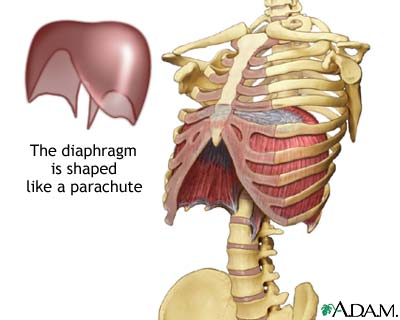
Movements of the thoracic wall generated by the actions of several muscles working together are responsible for respiration.  Muscles such as the intercostals, transversus thoracis, subcostals, scalenes, and the diaphragm have important roles in respiration.  These muscles work together to expand or compress the volume of the thoracic cavity by elevating and compressing the ribs, respectively.  There is a difference in the muscles involved in inspiration and expiration depending on whether it is passive or active.  During passive inspiration the primary muscles of inspiration are working, and these muscles are the diaphragm and the external intercostals.  During passive expiration, the elastic recoil due to the relaxation of the diaphragm and of the internal intercostals compresses the throacic cavity.  On the other hand, during active respiration more movement of the rib cage is necessary, so more muscles are involved.  During active inspiration the primary and secondary muscles are involved with the secondary being the sternocleidomastoid, scalenes, pectoralis major, pectoralis minor, serratus anterior, serratus posterior superior, and the upper iliocostalis.  Active expiration involves the recoil along with the abdominals, lower iliocostalis, lower longissimus, and the serratus posterior inferior.  The levatores costarum muscles are interesting in that they elevate the ribs, but it is not known whether or not they actually play a part in inspiration.


Pneumothorax is a somewhat common thoracic problem.  This occurs most often due to an injury, rib fracture, or following surgery and allows air to enter the pleural cavity, causing the lung to collapse.  If the pneumothorax is minor enough it may heal on its own, however, a needle is usually used to extract air from around the lung so that the lung will have better expansion. The needle is inserted into the 2nd intercostal space in the midclavicular line and into the pleural space.  The needle is inserted over the top of the 3rd rib to avoid the intercostal vessels and nerves.  For a larger pneumothorax a chest tube is required to extract the air.  The chest tube is inserted behind the pectoralis major in the mid-axillary line so that the thick muscle will not have to be dissected.  While keeping in mind that the diaphragm rises to the level of the 5th intercostal space during expiration, the highest intercostal space that can be felt is used for insertion of the chest tube.  Exact placement of the tube depends on the cause of the pneumothorax.

Site of Insertion

A. Robinson
Sources:
Moore, Dalley, Agur: Clinically Oriented Anatomy. 6th ed. Baltimore:  Lippincott Williams & Wilkins, 2010.
Gilroy, MacPherson, Ross:  Atlas of Anatomy. New York:  Thieme, 2008.
http://www.beltina.org/health-dictionary/diaphragm-breathing-function-definition.html
http://prepgmedicos.redstetho.com/forum/viewtopic.php?f=20&t=3150
http://anatomytopics.wordpress.com/category/thorax/

Thursday, March 24, 2011

The Hand & Wrist

I would like to touch on a few general topics relating to my specialty dissection of the hand.

The hand is considered to be the manual region of the upper limb distal to the forearm.  Twenty-seven bones constitute the hand and wrist and are considered as carpals, metacarpals, and phalanges.  There are eight carpal bones which are arranged in a proximal row and a distal row.  The proximal row includes the scaphoid, lunate, triquetrum, and pisiform bones while the distal rowincludes the trapezium, trapezoid, capitate, and hamate bones.  The distal row of the carpal bones articulate distally with the proximal surfaces of the metacarpals, also called the bases of the metacarpals.  There are five metacarpals, corresponding to digit 1 to digit 5, and articulating distally with the proximal phalanges, creating the knuckles of the hand.  There are fourteen phalanges total, which include the proximal, middle, and distal phalanges of digit 2 through digit 5.  Digit 1, the pollex, is made up of two phalanges, a proximal phalanx and a distal phalanx, rather than three.  These bones of the wrist and hand serve as anatomical landmarks as well as attachment points for the muscles that move the hand.

Many of the muscles of the forearm have tendons that insert on the hand including the:  flexor carpi radialis, palmaris longus, flexor carpi ulnaris, flexor digitorum superficialis, flexor digitorum profundis, flexor pollicis longus, extensor carpi radialis longus (ECRL), extensor carpi radialis brevis (ECRB), extensor digitorum, extensor digiti minimi (EDM), extensor carpi ulnaris, abductor pollicis longus (APL), extensor pollicis brevis (EPB), extensor pollicis longus (EPL), extensor indicis.  These muscles perform actions such as flexion, extension, adduction, and abduction of the hand as well as flexion and extension of the digits.  The tendons of these muscles pass under the extensor retinaculum and the flexor retinaculum, wrapped in synovial tendon sheaths, preventing the tendons from bowing and holding them in place on their osseofibrous tunnels.  On the dorsal side of the hand there are six synovial tendon sheaths and each lies in a different osseofibrous tunnel, so there are also six osseofibrous tunnels, and through these pass twelve tendons of nine extensor muscles.  The first tendon sheath includes the tendons for APL and EPB; the second tendon sheath includes the tendons of the ECRL and ECRB; the third tendon sheath includes the tendon of EPL; the fourth tendon sheath includes the tendon for extensor indicis as well as the four tendons of extensor digitorum; the fifth tendon sheath includes the tendon of EDM; the sixth tendon sheath includes the tendon of extensor carpi ulnaris.  In contrast to the numerous tendon sheaths on the dorsal side of the hand, on the palmar side there are only three:  a common sheath containing the tendons of flexor digitorum superficialis and flexor digitorum profundus; a sheath containing the tendon of flexor pollicis longus; a sheath containing flexor carpi radialis.

5 dorsal tendon compartments 


Adduction and abduction of the digits is primarily performed by the interosseous muscles of the hand, except for in the case of digit 1, the pollex.  The interosseous muscles insert on the extensor expansions which are flattened areas of four tendons found on the dorsal surface of the metacarpals as well as along the phalanges of digits 2-5.  The extensor expansions function to hold the extensor tendon in a central position along each of digits 2-5.  After passing through the extensor expansion, the extensor tendon divides into a median band and two lateral bands which continue to the base of the middle phalanx and to either side of the distal phalanx base.  The lateral bands provide insertion points for tendons of both the interosseous muscles and the lumbrical muscles of the hand.  To even further stabilize the extensor tendons, the extensor expansions are attached to the palmar ligament on both sides. 


On the palmar surface of the hand is an insertion point of the plamaris longus muscle, the palmar aponeurosis.  When the palmaris longus muscle is present, which is not always the case, the palmar aponeurosis is the expanded tendon of the muscle.  The palmar aponeurosis is the deep, and very thick, portion of the palmar fascia that distally extends to form four bands that attach to the bases of the proximal phalanges.  In contrast to the thickness of the palmar aponeurosis, there are two areas of the palmar fascia that are much thinner and these areas are known as the thenar eminence and the hypothenar eminence.  The thenar fascia covers the thenar eminence at the base of the pollex, whereas the hypothenar fascia covers the hypothenar eminence at the base of digit 5.  Somewhat between these two eminences is where the palmar aponeurosis can be found. 

The muscles that make up the thenar and hypothenar eminences are intrinsic muscles, meaning that their proximal attachment point is on the hand.  The muscles of the thenar eminence include the abductor pollicis brevis, flexor pollicis brevis, and the opponens pollicis, whereas the muscles of the hypothernar eminence include the abductor digiti minimi, flexor digiti minimi, and the opponens digiti minimi.  The thenar and hypothenar groups of muscles are often considered the thenar and hypothenar compartments, respectively.  When considering the compartments of intrinsic muscles of the hand, there are also three other compartments in addition to the thenar and hypothenar:  the adductor compartment, the central compartment, and the interosseous compartment.  In terms of the muscular contents of these other three compartments, the adductor compartment is made up of the adductor pollicis muscle, the central compartment includes the lumbrical muscles, and the interossei muscles are found in separate interosseous compartments.  Although only the muscular contents of the compartments are listed, the contents of the compartments do not consist of only muscles.

The thenar and hypothenar compartments are two of the more easily found landmarks of the surface anatomy of the hand.  The thenar eminence serves as a way to identify the location of the superficial and deep palmar arches.  The superficial palmar arch is found in the center of the palm,  terminating on the thenar eminence, and the deep palmar arch is located about 1cm more proximal than the superficial palmar arch.  Another anatomical landmark on the hand is the knuckle formed between the head of the metacarpal and the proximal phalanx.  The knuckles are important landmarks when finger amputations are performed.  While the aforementioned landmarks were not discussed in class for various reasons, the anatomical snuff box was an area that was heavily discussed.  The anatomical snuff box is an area which is medially bordered by the tendon of the extensor pollicis longus, and laterally bordered by the tendons of the extensor pollicis brevis and the tendon of the abductor pollicis longus.  This is an area used to palpate the scaphoid bone when it is thought to have possibly been fractured following a fall that was caught by bracing one's self with the hand.
Diagram of hand surface
A. Robinson
Sources:
Moore, Dalley, Agur: Clinically Oriented Anatomy- 6th Edition, 2010.